The Philippine journal of science. [Vol. 67, no. 1]
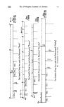
360 The Philippine Journal of Science 1938 Below the subopercle and mostly covered by it is the most posterior and outermost branchiostegal (Plate 2; text fig. 1, 39). There are four branchiostegals on each side, gradually diminishing in breadth and becoming more pointed mesially. They are all flat, thin, and lamellate. The two inner elements are attached to the lower half of the outer face of the ceratohyal (35), the two outer bones, to the same region in the epihyal. The elements of the opercular series grade off regularly into the subopercle and opercle, while the preopercle and interopercle seem not to fit in it. Hyobranchial series (Plates 2 and 5).-There is no mistaking the hyoid portion of the hyobranchial series, because it is the most anterior in position and much stouter in structure than the rest. The glossohyal (Plates 2 and 5, 37), forming the framework of the tongue, projects forward from the median region of the hyoid, immediately between the closely articulated two-layered hypohyals (36). It is mainly cartilaginous, but the posterior half is covered by a thin membrane bone which extends a little backward, covering the anterior portion of the 1st basibranchial. There are two sets of hypohyals (Plates 2 and 5, 36), one on top of the other. The dorsal parts are roughly circular in form and smaller in size than the ventral set. The ceratohyal (Plates 2 and 5, 35) extends backward and.outward from the hypohyals, followed still more posteriorly by the epihyal (Plates 2 and 5, 34) which is in length roughly only one-half the ceratohyal. The most posterior tip of the epihyal, a little nearer the dorsoposterior border, bears a small nodule of cartilage, the interhyal (Plates 2 and 5, 33), which articulates the ventral half of the hyoid arch with its dorsal half, the hyomandibular. The large urohyal (Plates 2 and 5, 38) articulates with the posterior median portion of the hypohyals. Posteriorly it comes in close contact with the anterior border of the clavicular parts of the pectoral girdle, in such an intimate manner that its dorsal ridge merges gracefully into the median ridge formed by the clavicles (56). There are three basibranchials (Plate 5, 40), the 1st and 2d of about equal size, and the 3d the largest of them all. A small flake of bone develops over the cartilage in the posterior half of the 1st basibranchial (Plate 5, 40,), partly covering the anterior portion of the 2d. The 2d basibranchial (Plate 5, 402) possesses a similar flake of bone in the same region as the 1st, which in
-
Scan 1

Page #1
-
Scan 2

Page #2
-
Scan 3

Page #3
-
Scan 4

Page #4
-
Scan 5

Page #5
-
Scan 6

Page #6
-
Scan 7

Page I - Title Page
-
Scan 8

Page II
-
Scan 9

Page III - Table of Contents
-
Scan 10

Page IV - Table of Contents
-
Scan 11

Page 1
-
Scan 12

Page 2
-
Scan 13

Page 3
-
Scan 14

Page 4
-
Scan 15

Page 5
-
Scan 16

Page 6
-
Scan 17

Page 7
-
Scan 18

Page 8
-
Scan 19

Page 9
-
Scan 20

Page 10
-
Scan 21

Page 11
-
Scan 22

Page 12
-
Scan 23

Page 13
-
Scan 24

Page 14
-
Scan 25

Page 15
-
Scan 26

Page 16
-
Scan 27

Page 17
-
Scan 28

Page 18
-
Scan 29

Page 19
-
Scan 30

Page 20
-
Scan 31

Page 21
-
Scan 32

Page 22
-
Scan 33

Page 23
-
Scan 34

Page 24
-
Scan 35

Page 25
-
Scan 36

Page 26
-
Scan 37

Page 27
-
Scan 38

Page 28
-
Scan 39

Page 29
-
Scan 40

Page 30
-
Scan 41
Page 31
-
Scan 42

Page 32
-
Scan 43

Page 33
-
Scan 44

Page 34
-
Scan 45

Page 35
-
Scan 46

Page 36
-
Scan 47

Page 37
-
Scan 48

Page 38
-
Scan 49

Page 39
-
Scan 50

Page 40
-
Scan 51

Page 41
-
Scan 52

Page 42
-
Scan 53

Page 43
-
Scan 54

Page 44
-
Scan 55

Page 45
-
Scan 56

Page 46
-
Scan 57

Page 47
-
Scan 58

Page 48
-
Scan 59

Page 49
-
Scan 60

Page 50
-
Scan 61

Page 51
-
Scan 62

Page 52
-
Scan 63

Page 53
-
Scan 64

Page 54
-
Scan 65

Page 55
-
Scan 66

Page 56
-
Scan 67

Page 57
-
Scan 68

Page 58
-
Scan 69

Page 59
-
Scan 70

Page 60
-
Scan 71

Page 61
-
Scan 72

Page 62
-
Scan 73

Page 63
-
Scan 74

Page 64
-
Scan 75

Page 65
-
Scan 76

Page 66
-
Scan 77

Page 67
-
Scan 78

Page 68
-
Scan 79
Page 69
-
Scan 80

Page 70
-
Scan 81

Page 71
-
Scan 82

Page 72
-
Scan 83

Page 73
-
Scan 84

Page 74
-
Scan 85

Page 75
-
Scan 86

Page 76
-
Scan 87

Page 77
-
Scan 88

Page 78
-
Scan 89

Page 79
-
Scan 90

Page 80
-
Scan 91

Page 81
-
Scan 92

Page 82
-
Scan 93

Page 83
-
Scan 94

Page 84
-
Scan 95

Page 85
-
Scan 96

Page 86
-
Scan 97

Page 87
-
Scan 98

Page 88
-
Scan 99

Page 89
-
Scan 100

Page 90
-
Scan 101

Page 91
-
Scan 102

Page 92
-
Scan 103

Page 93
-
Scan 104

Page 94
-
Scan 105

Page 95
-
Scan 106

Page 96
-
Scan 107

Page 97
-
Scan 108

Page 98
-
Scan 109

Page 99
-
Scan 110

Page 100
-
Scan 111

Page 101
-
Scan 112

Page 102
-
Scan 113

Page 103
-
Scan 114

Page 104
-
Scan 115

Page 105
-
Scan 116

Page 106
-
Scan 117

Page 107
-
Scan 118

Page 108
-
Scan 119

Page 109
-
Scan 120

Page 110
-
Scan 121

Page #121
-
Scan 122

Page #122
-
Scan 123

Page #123
-
Scan 124

Page #124
-
Scan 125

Page #125
-
Scan 126

Page #126
-
Scan 127

Page #127
-
Scan 128

Page #128
-
Scan 129

Page #129
-
Scan 130

Page #130
-
Scan 131

Page #131
-
Scan 132

Page #132
-
Scan 133

Page #133
-
Scan 134

Page #134
-
Scan 135

Page #135
-
Scan 136

Page #136
-
Scan 137
Page #137
-
Scan 138

Page #138
-
Scan 139

Page #139
-
Scan 140

Page #140
-
Scan 141

Page #141
-
Scan 142

Page #142
-
Scan 143

Page 111
-
Scan 144

Page 112
-
Scan 145

Page 113
-
Scan 146

Page 114
-
Scan 147

Page 115
-
Scan 148

Page 116
-
Scan 149

Page 117
-
Scan 150

Page 118
-
Scan 151

Page 119
-
Scan 152

Page 120
-
Scan 153

Page 121
-
Scan 154

Page 122
-
Scan 155

Page 123
-
Scan 156

Page 124
-
Scan 157

Page 125
-
Scan 158

Page 126
-
Scan 159

Page 127
-
Scan 160

Page 128
-
Scan 161
Page 129
-
Scan 162

Page 130
-
Scan 163

Page 131
-
Scan 164

Page 132
-
Scan 165

Page 133
-
Scan 166

Page 134
-
Scan 167

Page 135
-
Scan 168

Page 136
-
Scan 169

Page 137
-
Scan 170

Page 138
-
Scan 171

Page 139
-
Scan 172

Page 140
-
Scan 173

Page 141
-
Scan 174

Page 142
-
Scan 175

Page 143
-
Scan 176

Page 144
-
Scan 177

Page 145
-
Scan 178

Page 146
-
Scan 179

Page 147
-
Scan 180

Page 148
-
Scan 181

Page 149
-
Scan 182

Page 150
-
Scan 183

Page 151
-
Scan 184

Page 152
-
Scan 185

Page 153
-
Scan 186

Page 154
-
Scan 187

Page 155
-
Scan 188

Page 156
-
Scan 189

Page 157
-
Scan 190

Page 158
-
Scan 191

Page 159
-
Scan 192

Page 160
-
Scan 193

Page 161
-
Scan 194

Page 162
-
Scan 195

Page 163
-
Scan 196

Page 164
-
Scan 197

Page 165
-
Scan 198

Page 166
-
Scan 199

Page #199
-
Scan 200

Page #200
-
Scan 201

Page #201
-
Scan 202

Page #202
-
Scan 203

Page #203
-
Scan 204

Page #204
-
Scan 205
Page 167
-
Scan 206

Page 168
-
Scan 207

Page 169
-
Scan 208

Page 170
-
Scan 209

Page 171
-
Scan 210

Page 172
-
Scan 211

Page 173
-
Scan 212

Page 174
-
Scan 213

Page 175
-
Scan 214

Page 176
-
Scan 215

Page 177
-
Scan 216

Page 178
-
Scan 217

Page 179
-
Scan 218

Page 180
-
Scan 219

Page 181
-
Scan 220

Page 182
-
Scan 221

Page 183
-
Scan 222

Page 184
-
Scan 223

Page 185
-
Scan 224

Page 186
-
Scan 225

Page 187
-
Scan 226

Page 188
-
Scan 227

Page #227
-
Scan 228

Page #228
-
Scan 229

Page #229
-
Scan 230

Page #230
-
Scan 231

Page 189
-
Scan 232

Page 190
-
Scan 233

Page 191
-
Scan 234

Page 192
-
Scan 235

Page 193
-
Scan 236

Page 194
-
Scan 237

Page 195
-
Scan 238
Page 196
-
Scan 239

Page 197
-
Scan 240

Page 198
-
Scan 241

Page 199
-
Scan 242

Page 200
-
Scan 243

Page #243
-
Scan 244

Page #244
-
Scan 245

Page #245
-
Scan 246

Page #246
-
Scan 247

Page #247
-
Scan 248

Page #248
-
Scan 249

Page #249
-
Scan 250

Page 201
-
Scan 251

Page 202
-
Scan 252

Page 203
-
Scan 253

Page 204
-
Scan 254

Page 205
-
Scan 255

Page 206
-
Scan 256

Page #256
-
Scan 257

Page #257
-
Scan 258

Page 207
-
Scan 259

Page 208
-
Scan 260

Page 209
-
Scan 261

Page 210
-
Scan 262

Page 211
-
Scan 263

Page 212
-
Scan 264

Page 213
-
Scan 265

Page 214
-
Scan 266

Page 215
-
Scan 267

Page 216
-
Scan 268

Page 217
-
Scan 269

Page 218
-
Scan 270

Page 219
-
Scan 271

Page 220
-
Scan 272

Page 221
-
Scan 273

Page 222
-
Scan 274

Page 223
-
Scan 275

Page 224
-
Scan 276

Page 225
-
Scan 277
Page 226
-
Scan 278

Page 227
-
Scan 279

Page 228
-
Scan 280

Page 229
-
Scan 281

Page 230
-
Scan 282

Page #282
-
Scan 283

Page #283
-
Scan 284

Page #284
-
Scan 285

Page #285
-
Scan 286

Page #286
-
Scan 287

Page #287
-
Scan 288

Page #288
-
Scan 289

Page #289
-
Scan 290

Page #290
-
Scan 291

Page #291
-
Scan 292

Page 231
-
Scan 293

Page 232
-
Scan 294

Page 233
-
Scan 295

Page 234
-
Scan 296

Page 235
-
Scan 297

Page 236
-
Scan 298

Page 237
-
Scan 299

Page 238
-
Scan 300

Page 239
-
Scan 301

Page 240
-
Scan 302

Page 241
-
Scan 303

Page 242
-
Scan 304

Page 243
-
Scan 305

Page 244
-
Scan 306

Page 245
-
Scan 307
Page 246
-
Scan 308

Page 247
-
Scan 309

Page 248
-
Scan 310

Page 249
-
Scan 311

Page 250
-
Scan 312

Page 251
-
Scan 313

Page 252
-
Scan 314

Page 253
-
Scan 315

Page 254
-
Scan 316

Page 255
-
Scan 317

Page 256
-
Scan 318

Page 257
-
Scan 319

Page 258
-
Scan 320

Page 259
-
Scan 321

Page 260
-
Scan 322

Page 261
-
Scan 323

Page 262
-
Scan 324

Page 263
-
Scan 325

Page 264
-
Scan 326

Page 265
-
Scan 327

Page 266
-
Scan 328

Page 267
-
Scan 329

Page 268
-
Scan 330

Page 269
-
Scan 331

Page 270
-
Scan 332

Page 271
-
Scan 333

Page 272
-
Scan 334

Page 273
-
Scan 335

Page 274
-
Scan 336

Page 275
-
Scan 337

Page 276
-
Scan 338

Page 277
-
Scan 339

Page 278
-
Scan 340

Page 279
-
Scan 341

Page 280
-
Scan 342

Page 281
-
Scan 343

Page 282
-
Scan 344

Page 283
-
Scan 345

Page 284
-
Scan 346

Page 285
-
Scan 347

Page 286
-
Scan 348

Page 287
-
Scan 349

Page 288
-
Scan 350

Page 289
-
Scan 351

Page 290
-
Scan 352

Page 291
-
Scan 353

Page 292
-
Scan 354

Page 293
-
Scan 355

Page 294
-
Scan 356

Page 295
-
Scan 357

Page 296
-
Scan 358

Page 297
-
Scan 359

Page 298
-
Scan 360

Page 299
-
Scan 361

Page 300
-
Scan 362

Page 301
-
Scan 363

Page 302
-
Scan 364

Page 303
-
Scan 365

Page 304
-
Scan 366

Page 305
-
Scan 367

Page 306
-
Scan 368

Page 307
-
Scan 369

Page 308
-
Scan 370

Page 309
-
Scan 371

Page 310
-
Scan 372

Page 311
-
Scan 373

Page 312
-
Scan 374

Page 313
-
Scan 375

Page 314
-
Scan 376

Page 315
-
Scan 377

Page 316
-
Scan 378

Page 317
-
Scan 379

Page 318
-
Scan 380

Page 319
-
Scan 381

Page 320
-
Scan 382
Page 321
-
Scan 383

Page 322
-
Scan 384

Page 323
-
Scan 385

Page 324
-
Scan 386

Page 325
-
Scan 387

Page 326
-
Scan 388

Page 327
-
Scan 389

Page 328
-
Scan 390

Page 329
-
Scan 391

Page 330
-
Scan 392

Page 331
-
Scan 393

Page 332
-
Scan 394

Page 333
-
Scan 395

Page 334
-
Scan 396

Page #396
-
Scan 397

Page #397
-
Scan 398

Page #398
-
Scan 399

Page #399
-
Scan 400

Page #400
-
Scan 401

Page #401
-
Scan 402

Page #402
-
Scan 403

Page #403
-
Scan 404

Page #404
-
Scan 405

Page 335
-
Scan 406

Page 336
-
Scan 407

Page 337
-
Scan 408

Page 338
-
Scan 409

Page 339
-
Scan 410

Page 340
-
Scan 411

Page 341
-
Scan 412

Page 342
-
Scan 413

Page 343
-
Scan 414

Page 344
-
Scan 415

Page 345
-
Scan 416

Page 346
-
Scan 417

Page 347
-
Scan 418

Page 348
-
Scan 419

Page 349
-
Scan 420

Page 350
-
Scan 421
Page 351
-
Scan 422

Page 352
-
Scan 423

Page 353
-
Scan 424

Page 354
-
Scan 425

Page 355
-
Scan 426

Page 356
-
Scan 427

Page 357
-
Scan 428

Page 358
-
Scan 429

Page 359
-
Scan 430

Page 360
-
Scan 431

Page 361
-
Scan 432

Page 362
-
Scan 433

Page 363
-
Scan 434

Page 364
-
Scan 435
Page 365
-
Scan 436

Page 366
-
Scan 437

Page 367
-
Scan 438

Page 368
-
Scan 439

Page 369
-
Scan 440

Page 370
-
Scan 441

Page 371
-
Scan 442

Page 372
-
Scan 443

Page 373
-
Scan 444

Page 374
-
Scan 445

Page 375
-
Scan 446

Page 376
-
Scan 447

Page 377
-
Scan 448

Page 378
-
Scan 449

Page #449
-
Scan 450

Page #450
-
Scan 451

Page #451
-
Scan 452

Page #452
-
Scan 453

Page #453
-
Scan 454

Page #454
-
Scan 455

Page #455
-
Scan 456

Page #456
-
Scan 457

Page #457
-
Scan 458

Page #458
-
Scan 459

Page 379
-
Scan 460

Page 380
-
Scan 461

Page 381
-
Scan 462

Page 382
-
Scan 463

Page 383
-
Scan 464

Page 384
-
Scan 465

Page 385
-
Scan 466

Page 386
-
Scan 467

Page 387
-
Scan 468

Page 388
-
Scan 469

Page 389
-
Scan 470

Page 390
-
Scan 471

Page 391
-
Scan 472

Page 392
-
Scan 473

Page #473
-
Scan 474

Page #474
-
Scan 475

Page #475
-
Scan 476

Page #476
-
Scan 477

Page #477
-
Scan 478

Page #478
-
Scan 479

Page #479
-
Scan 480

Page #480
-
Scan 481

Page #481
-
Scan 482

Page #482
-
Scan 483

Page 393
-
Scan 484

Page 394
-
Scan 485

Page 395
-
Scan 486
Page 396
-
Scan 487

Page 397
-
Scan 488

Page 398
-
Scan 489

Page #489
-
Scan 490

Page #490
-
Scan 491

Page #491
-
Scan 492

Page #492
-
Scan 493

Page 399
-
Scan 494

Page 400
-
Scan 495

Page 401
-
Scan 496

Page 402
-
Scan 497

Page 403
-
Scan 498

Page 404
-
Scan 499

Page 405
-
Scan 500

Page 406
-
Scan 501

Page 407
-
Scan 502

Page 408
-
Scan 503

Page 409
-
Scan 504

Page #504
-
Scan 505

Page 411
-
Scan 506

Page 412
-
Scan 507

Page 413
-
Scan 508

Page 414
-
Scan 509

Page 415
-
Scan 510

Page 416
-
Scan 511

Page 417
-
Scan 512

Page 418
-
Scan 513

Page 419
-
Scan 514

Page 420
-
Scan 515

Page #515
-
Scan 516

Page #516
-
Scan 517

Page 421
-
Scan 518

Page 422
-
Scan 519

Page 423
-
Scan 520

Page 424
-
Scan 521

Page 425
-
Scan 522

Page 426
-
Scan 523

Page 427
-
Scan 524

Page 428
-
Scan 525

Page 429
-
Scan 526

Page 430
-
Scan 527

Page 431
-
Scan 528

Page 432
-
Scan 529

Page 433
-
Scan 530

Page 434
-
Scan 531

Page 435
-
Scan 532

Page 436
-
Scan 533

Page 437 - Comprehensive Index
-
Scan 534

Page 438 - Comprehensive Index
-
Scan 535

Page 439 - Comprehensive Index
-
Scan 536

Page 440 - Comprehensive Index
-
Scan 537

Page 441 - Comprehensive Index
-
Scan 538

Page 442 - Comprehensive Index
-
Scan 539

Page 443 - Comprehensive Index
-
Scan 540

Page 444 - Comprehensive Index
-
Scan 541

Page 445 - Comprehensive Index
-
Scan 542

Page 446 - Comprehensive Index
-
Scan 543

Page 447 - Comprehensive Index
-
Scan 544

Page 448 - Comprehensive Index
-
Scan 545

Page 449 - Comprehensive Index
-
Scan 546

Page 450 - Comprehensive Index
-
Scan 547

Page 451 - Comprehensive Index
-
Scan 548

Page 452 - Comprehensive Index
-
Scan 549

Page 453 - Comprehensive Index
-
Scan 550

Page 454 - Comprehensive Index
-
Scan 551

Page 455 - Comprehensive Index
-
Scan 552

Page 456 - Comprehensive Index
-
Scan 553

Page 457 - Comprehensive Index
-
Scan 554

Page 458 - Comprehensive Index
-
Scan 555

Page #555 - Title Page
-
Scan 556

Page #556
-
Scan 557

Page #557
-
Scan 558

Page #558 - Table of Contents
-
Scan 559

Page #559
-
Scan 560

Page #560
-
Scan 561

Page #561
-
Scan 562

Page #562
-
Scan 563

Page #563
-
Scan 564

Page #564
About this Item
- Title
- The Philippine journal of science. [Vol. 67, no. 1]
- Canvas
- Page 360
- Publication
- Manila: Philippines Bureau of Science,
- 1906-
- Subject terms
- Science -- Periodicals
Technical Details
- Link to this Item
-
https://name.umdl.umich.edu/act3868.0067.001
- Link to this scan
-
https://quod.lib.umich.edu/p/philamer/act3868.0067.001/430
Rights and Permissions
The University of Michigan Library provides access to these materials for educational and research purposes. These materials may be under copyright. If you decide to use any of these materials, you are responsible for making your own legal assessment and securing any necessary permission.
Related Links
IIIF
- Manifest
-
https://quod.lib.umich.edu/cgi/t/text/api/manifest/philamer:act3868.0067.001
Cite this Item
- Full citation
-
"The Philippine journal of science. [Vol. 67, no. 1]." In the digital collection The United States and its Territories, 1870 - 1925: The Age of Imperialism. https://name.umdl.umich.edu/act3868.0067.001. University of Michigan Library Digital Collections. Accessed June 13, 2025.