The boletes of Michigan, by Alexander H. Smith and Harry D. Thiers.
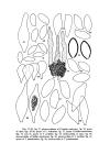
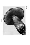
INTRODUCTION 11 usually become shorter and branching occurs to give rise to the hymenial elements. The diverging hyphae in most boletes are somewhat gelatinous, which causes the whole hymenophore, to some extent, to have a similar consistency. Because the texture is very pliant, good freehand sections of the tube layer are difficult to obtain. Also, the degree to which the individual hyphae are separated from each other, or the angle at which they diverge, is likely to be variable for a number of reasons. First, the more gelatinized the hyphae become the farther apart from each other they are separated in the trama because of the swelling effect the intervening slime exerts. Second, if sections are made from material killed and fixed in such solutions as FAA, one cannot be sure the picture one gets actually represents the arrangement in the living material even though the technique always gives the same result. Sections may be cut on a freezing microtome, but here again the freezing may very well effect the degree of gelatinization of the hyphae. Finally, the differences, even when shown to best advantage, are not sufficient to be of material aid in distinguishing genera or species in the family. The feature of bilateral hymenophoral trama is most valuable as a character to aid in distinguishing the Boletaceae as a family-especially from the true polypores. In the trama of the basidiocarp, in addition to the hyphae which form the basic tissue (matrical hyphae), one often observes hyphae distinguished in some way by a content different from that of the matrical hyphae. The content is often in the form of globular material or a dense homogeneous material which refracts the light differently than do the other hyphae. Collectively, such hyphae are regarded as a laticiferous system. We regard them as repository hyphae containing, possibly, by-products of metabolism, and the system is best regarded as a storage system rather than a system of "vascular hyphae" (which implies the function of conduction). The degree of their specialization in the boletes varies. Smith has seen the ends of some actually forming basidia with spores attached. Rarely in the boletes do they actually contain a latex that is exuded on injured places of the basidiocarp or on the developing pore surface of the hymenophore. THE STRUCTURE OF THE PILEUS For taxonomic purposes the dermal layer of the pileus is the most important and will be treated first. The important characters are in the arrangement of the hyphae and, second, in their anatomical features.
-
Scan 1

Page #1
-
Scan 2

Page #2
-
Scan 3

Page #3 - Title Page
-
Scan 4

Page #4
-
Scan 5

Page #5
-
Scan 6

Page #6
-
Scan 7

Page #7 - Table of Contents
-
Scan 8

Page #8
-
Scan 9

Page 1
-
Scan 10

Page 2
-
Scan 11

Page 3
-
Scan 12

Page 4
-
Scan 13

Page 5
-
Scan 14

Page 6
-
Scan 15

Page 7
-
Scan 16

Page 8
-
Scan 17

Page 9
-
Scan 18

Page 10
-
Scan 19

Page 11
-
Scan 20

Page 12
-
Scan 21

Page 13
-
Scan 22

Page 14
-
Scan 23

Page 15
-
Scan 24

Page 16
-
Scan 25

Page 17
-
Scan 26

Page 18
-
Scan 27

Page 19
-
Scan 28

Page 20
-
Scan 29

Page 21
-
Scan 30

Page 22
-
Scan 31

Page 23
-
Scan 32

Page 24
-
Scan 33

Page 25
-
Scan 34

Page 26
-
Scan 35

Page 27
-
Scan 36

Page 28
-
Scan 37

Page 29
-
Scan 38

Page 30
-
Scan 39

Page 31
-
Scan 40

Page 32
-
Scan 41

Page 33
-
Scan 42

Page 34
-
Scan 43

Page 35
-
Scan 44

Page 36
-
Scan 45

Page 37
-
Scan 46

Page 38
-
Scan 47

Page 39
-
Scan 48

Page 40
-
Scan 49

Page 41
-
Scan 50

Page 42
-
Scan 51

Page 43
-
Scan 52

Page 44
-
Scan 53

Page 45
-
Scan 54

Page 46
-
Scan 55

Page 47
-
Scan 56

Page 48
-
Scan 57

Page 49
-
Scan 58

Page 50
-
Scan 59

Page 51
-
Scan 60

Page 52
-
Scan 61

Page 53
-
Scan 62

Page 54
-
Scan 63

Page 55
-
Scan 64

Page 56
-
Scan 65

Page 57
-
Scan 66

Page 58
-
Scan 67

Page 59
-
Scan 68

Page 60
-
Scan 69

Page 61
-
Scan 70

Page 62
-
Scan 71

Page 63
-
Scan 72

Page 64
-
Scan 73

Page 65
-
Scan 74

Page 66
-
Scan 75

Page 67
-
Scan 76

Page 68
-
Scan 77

Page 69
-
Scan 78

Page 70
-
Scan 79

Page 71
-
Scan 80

Page 72
-
Scan 81

Page 73
-
Scan 82

Page 74
-
Scan 83

Page 75
-
Scan 84

Page 76
-
Scan 85

Page 77
-
Scan 86

Page 78
-
Scan 87

Page 79
-
Scan 88

Page 80
-
Scan 89

Page 81
-
Scan 90

Page 82
-
Scan 91

Page 83
-
Scan 92

Page 84
-
Scan 93

Page 85
-
Scan 94

Page 86
-
Scan 95

Page 87
-
Scan 96

Page 88
-
Scan 97

Page 89
-
Scan 98

Page 90
-
Scan 99

Page 91
-
Scan 100

Page 92
-
Scan 101

Page 93
-
Scan 102

Page 94
-
Scan 103

Page 95
-
Scan 104

Page 96
-
Scan 105

Page 97
-
Scan 106

Page 98
-
Scan 107

Page 99
-
Scan 108

Page 100
-
Scan 109

Page 101
-
Scan 110

Page 102
-
Scan 111

Page 103
-
Scan 112

Page 104
-
Scan 113

Page 105
-
Scan 114
Page 106
-
Scan 115

Page 107
-
Scan 116

Page 108
-
Scan 117

Page 109
-
Scan 118

Page 110
-
Scan 119

Page 111
-
Scan 120

Page 112
-
Scan 121

Page 113
-
Scan 122

Page 114
-
Scan 123

Page 115
-
Scan 124

Page 116
-
Scan 125

Page 117
-
Scan 126

Page 118
-
Scan 127

Page 119
-
Scan 128

Page 120
-
Scan 129

Page 121
-
Scan 130

Page 122
-
Scan 131

Page 123
-
Scan 132

Page 124
-
Scan 133

Page 125
-
Scan 134

Page 126
-
Scan 135

Page 127
-
Scan 136

Page 128
-
Scan 137

Page 129
-
Scan 138

Page 130
-
Scan 139

Page 131
-
Scan 140

Page 132
-
Scan 141

Page 133
-
Scan 142

Page 134
-
Scan 143

Page 135
-
Scan 144

Page 136
-
Scan 145

Page 137
-
Scan 146

Page 138
-
Scan 147

Page 139
-
Scan 148

Page 140
-
Scan 149

Page 141
-
Scan 150

Page 142
-
Scan 151

Page 143
-
Scan 152

Page 144
-
Scan 153

Page 145
-
Scan 154

Page 146
-
Scan 155

Page 147
-
Scan 156

Page 148
-
Scan 157

Page 149
-
Scan 158

Page 150
-
Scan 159

Page 151
-
Scan 160

Page 152
-
Scan 161

Page 153
-
Scan 162

Page 154
-
Scan 163

Page 155
-
Scan 164

Page 156
-
Scan 165

Page 157
-
Scan 166

Page 158
-
Scan 167

Page 159
-
Scan 168

Page 160
-
Scan 169

Page 161
-
Scan 170

Page 162
-
Scan 171

Page 163
-
Scan 172

Page 164
-
Scan 173

Page 165
-
Scan 174

Page 166
-
Scan 175

Page 167
-
Scan 176

Page 168
-
Scan 177

Page 169
-
Scan 178

Page 170
-
Scan 179

Page 171
-
Scan 180

Page 172
-
Scan 181

Page 173
-
Scan 182

Page 174
-
Scan 183

Page 175
-
Scan 184

Page 176
-
Scan 185

Page 177
-
Scan 186

Page 178
-
Scan 187

Page 179
-
Scan 188

Page 180
-
Scan 189

Page 181
-
Scan 190

Page 182
-
Scan 191

Page 183
-
Scan 192

Page 184
-
Scan 193

Page 185
-
Scan 194

Page 186
-
Scan 195

Page 187
-
Scan 196

Page 188
-
Scan 197

Page 189
-
Scan 198

Page 190
-
Scan 199

Page 191
-
Scan 200

Page 192
-
Scan 201

Page 193
-
Scan 202

Page 194
-
Scan 203

Page 195
-
Scan 204

Page 196
-
Scan 205

Page 197
-
Scan 206

Page 198
-
Scan 207

Page 199
-
Scan 208

Page 200
-
Scan 209

Page 201
-
Scan 210

Page 202
-
Scan 211

Page 203
-
Scan 212

Page 204
-
Scan 213

Page 205
-
Scan 214

Page 206
-
Scan 215

Page 207
-
Scan 216

Page 208
-
Scan 217

Page 209
-
Scan 218

Page 210
-
Scan 219

Page 211
-
Scan 220

Page 212
-
Scan 221

Page 213
-
Scan 222

Page 214
-
Scan 223

Page 215
-
Scan 224

Page 216
-
Scan 225

Page 217
-
Scan 226

Page 218
-
Scan 227

Page 219
-
Scan 228

Page 220
-
Scan 229

Page 221
-
Scan 230

Page 222
-
Scan 231

Page 223
-
Scan 232

Page 224
-
Scan 233

Page 225
-
Scan 234

Page 226
-
Scan 235

Page 227
-
Scan 236

Page 228
-
Scan 237

Page 229
-
Scan 238

Page 230
-
Scan 239

Page 231
-
Scan 240

Page 232
-
Scan 241

Page 233
-
Scan 242

Page 234
-
Scan 243

Page 235
-
Scan 244

Page 236
-
Scan 245

Page 237
-
Scan 246

Page 238
-
Scan 247

Page 239
-
Scan 248

Page 240
-
Scan 249

Page 241
-
Scan 250

Page 242
-
Scan 251

Page 243
-
Scan 252

Page 244
-
Scan 253

Page 245
-
Scan 254

Page 246
-
Scan 255

Page 247
-
Scan 256

Page 248
-
Scan 257

Page 249
-
Scan 258

Page 250
-
Scan 259

Page 251
-
Scan 260

Page 252
-
Scan 261

Page 253
-
Scan 262

Page 254
-
Scan 263

Page 255
-
Scan 264

Page 256
-
Scan 265

Page 257
-
Scan 266

Page 258
-
Scan 267

Page 259
-
Scan 268

Page 260
-
Scan 269

Page 261
-
Scan 270

Page 262
-
Scan 271

Page 263
-
Scan 272

Page 264
-
Scan 273

Page 265
-
Scan 274

Page 266
-
Scan 275

Page 267
-
Scan 276

Page 268
-
Scan 277

Page 269
-
Scan 278

Page 270
-
Scan 279

Page 271
-
Scan 280

Page 272
-
Scan 281

Page 273
-
Scan 282

Page 274
-
Scan 283

Page 275
-
Scan 284

Page 276
-
Scan 285

Page 277
-
Scan 286

Page 278
-
Scan 287

Page 279
-
Scan 288

Page 280
-
Scan 289

Page 281
-
Scan 290

Page 282
-
Scan 291

Page 283
-
Scan 292

Page 284
-
Scan 293

Page 285
-
Scan 294

Page 286
-
Scan 295

Page 287
-
Scan 296

Page 288
-
Scan 297

Page 289
-
Scan 298

Page 290
-
Scan 299

Page 291
-
Scan 300

Page 292
-
Scan 301

Page 293
-
Scan 302

Page 294
-
Scan 303

Page 295
-
Scan 304

Page 296
-
Scan 305

Page 297
-
Scan 306

Page 298
-
Scan 307

Page 299
-
Scan 308

Page 300
-
Scan 309

Page 301
-
Scan 310

Page 302
-
Scan 311

Page 303
-
Scan 312

Page 304
-
Scan 313

Page 305
-
Scan 314

Page 306
-
Scan 315

Page 307
-
Scan 316

Page 308
-
Scan 317

Page 309
-
Scan 318

Page 310
-
Scan 319

Page 311
-
Scan 320

Page 312
-
Scan 321

Page 313
-
Scan 322

Page 314
-
Scan 323

Page 315
-
Scan 324

Page 316
-
Scan 325

Page 317
-
Scan 326

Page 318
-
Scan 327

Page 319
-
Scan 328

Page 320
-
Scan 329

Page 321
-
Scan 330

Page 322
-
Scan 331

Page 323
-
Scan 332

Page 324
-
Scan 333

Page 325
-
Scan 334

Page 326
-
Scan 335

Page 327
-
Scan 336

Page 328
-
Scan 337

Page 329
-
Scan 338

Page 330
-
Scan 339

Page 331
-
Scan 340

Page 332
-
Scan 341

Page 333
-
Scan 342

Page 334
-
Scan 343

Page 335
-
Scan 344

Page 336
-
Scan 345

Page 337
-
Scan 346

Page 338
-
Scan 347

Page 339
-
Scan 348

Page 340
-
Scan 349

Page 341
-
Scan 350

Page 342
-
Scan 351

Page 343
-
Scan 352

Page 344
-
Scan 353

Page 345
-
Scan 354

Page 346
-
Scan 355

Page 347
-
Scan 356

Page 348
-
Scan 357

Page 349
-
Scan 358

Page 350
-
Scan 359

Page 351
-
Scan 360

Page 352
-
Scan 361

Page 353
-
Scan 362

Page 354
-
Scan 363

Page 355
-
Scan 364

Page 356
-
Scan 365

Page 357
-
Scan 366

Page 358
-
Scan 367

Page 359
-
Scan 368

Page 360
-
Scan 369

Page 361
-
Scan 370

Page 362
-
Scan 371

Page 363
-
Scan 372

Page 364
-
Scan 373

Page 365
-
Scan 374

Page 366
-
Scan 375

Page 367
-
Scan 376

Page 368
-
Scan 377

Page 369
-
Scan 378

Page 370
-
Scan 379

Page 371
-
Scan 380
Page 372
-
Scan 381

Page 373
-
Scan 382

Page 374
-
Scan 383

Page 375
-
Scan 384

Page 376
-
Scan 385

Page 377
-
Scan 386

Page 378
-
Scan 387

Page 379
-
Scan 388

Page 380
-
Scan 389

Page 381
-
Scan 390

Page 382
-
Scan 391

Page 383
-
Scan 392

Page 384
-
Scan 393

Page 385
-
Scan 394

Page 386
-
Scan 395

Page 387
-
Scan 396

Page 388
-
Scan 397

Page 389
-
Scan 398

Page 390
-
Scan 399

Page 391
-
Scan 400

Page 392
-
Scan 401

Page 393
-
Scan 402

Page 394
-
Scan 403

Page 395
-
Scan 404

Page 396
-
Scan 405

Page 397
-
Scan 406

Page 398
-
Scan 407

Page 399
-
Scan 408

Page 400
-
Scan 409

Page 401
-
Scan 410

Page 402
-
Scan 411

Page 403
-
Scan 412

Page 404
-
Scan 413

Page 405
-
Scan 414

Page 406
-
Scan 415

Page 407
-
Scan 416

Page 408
-
Scan 417

Page 409
-
Scan 418

Page 410
-
Scan 419

Page 411
-
Scan 420

Page 412
-
Scan 421

Page 413
-
Scan 422

Page 414
-
Scan 423

Page 415
-
Scan 424

Page 416
-
Scan 425

Page 417
-
Scan 426

Page #426
-
Scan 427

Page 418
-
Scan 428

Page 419
-
Scan 429

Page #429
-
Scan 430

Page #430
-
Scan 431
Page #431
-
Scan 432

Page #432
-
Scan 433

Page #433
-
Scan 434

Page #434
-
Scan 435

Page #435
-
Scan 436

Page #436
-
Scan 437

Page #437
-
Scan 438

Page #438
-
Scan 439

Page #439
-
Scan 440

Page #440
-
Scan 441

Page #441
-
Scan 442

Page #442
-
Scan 443

Page #443
-
Scan 444

Page #444
-
Scan 445

Page #445
-
Scan 446

Page #446
-
Scan 447

Page #447
-
Scan 448

Page #448
-
Scan 449

Page #449
-
Scan 450
Page #450
-
Scan 451

Page #451
-
Scan 452

Page #452
-
Scan 453

Page #453
-
Scan 454

Page #454
-
Scan 455

Page #455
-
Scan 456

Page #456
-
Scan 457

Page #457
-
Scan 458

Page #458
-
Scan 459

Page #459
-
Scan 460

Page #460
-
Scan 461

Page #461
-
Scan 462

Page #462
-
Scan 463

Page #463
-
Scan 464

Page #464
-
Scan 465

Page #465
-
Scan 466

Page #466
-
Scan 467

Page #467
-
Scan 468

Page #468
-
Scan 469

Page #469
-
Scan 470

Page #470
-
Scan 471

Page #471
-
Scan 472

Page #472
-
Scan 473

Page #473
-
Scan 474

Page #474
-
Scan 475

Page #475
-
Scan 476

Page #476
-
Scan 477

Page #477
-
Scan 478

Page #478
-
Scan 479

Page #479
-
Scan 480

Page #480
-
Scan 481

Page #481
-
Scan 482

Page #482
-
Scan 483

Page #483
-
Scan 484

Page #484
-
Scan 485

Page #485
-
Scan 486

Page #486
-
Scan 487

Page #487
-
Scan 488

Page #488
-
Scan 489

Page #489
-
Scan 490

Page #490
-
Scan 491

Page #491
-
Scan 492

Page #492
-
Scan 493

Page #493
-
Scan 494

Page #494
-
Scan 495

Page #495
-
Scan 496

Page #496
-
Scan 497

Page #497
-
Scan 498

Page #498
-
Scan 499

Page #499
-
Scan 500

Page #500
-
Scan 501

Page #501
-
Scan 502

Page #502
-
Scan 503

Page #503
-
Scan 504

Page #504
-
Scan 505

Page #505
-
Scan 506

Page #506
-
Scan 507

Page #507
-
Scan 508

Page #508
-
Scan 509

Page #509
-
Scan 510

Page #510
-
Scan 511

Page #511
-
Scan 512

Page #512
-
Scan 513

Page #513
-
Scan 514

Page #514
-
Scan 515

Page #515
-
Scan 516

Page #516
-
Scan 517

Page #517
-
Scan 518

Page #518
-
Scan 519

Page #519
-
Scan 520

Page #520
-
Scan 521
Page #521
-
Scan 522

Page #522
-
Scan 523

Page #523
-
Scan 524

Page #524
-
Scan 525

Page #525
-
Scan 526

Page #526
-
Scan 527

Page #527
-
Scan 528

Page #528
-
Scan 529

Page #529
-
Scan 530

Page #530
-
Scan 531

Page #531
-
Scan 532

Page #532
-
Scan 533

Page #533
-
Scan 534

Page #534
-
Scan 535

Page #535
-
Scan 536

Page #536
-
Scan 537

Page #537
-
Scan 538

Page #538
-
Scan 539

Page #539
-
Scan 540

Page #540
-
Scan 541

Page #541
-
Scan 542

Page #542
-
Scan 543

Page #543
-
Scan 544

Page #544
-
Scan 545

Page #545
-
Scan 546

Page #546
-
Scan 547

Page #547
-
Scan 548

Page #548
-
Scan 549

Page #549
-
Scan 550

Page #550
-
Scan 551

Page #551
-
Scan 552

Page #552
-
Scan 553

Page #553
-
Scan 554

Page #554
-
Scan 555

Page #555
-
Scan 556

Page #556
-
Scan 557

Page #557
-
Scan 558

Page #558
-
Scan 559

Page #559
-
Scan 560

Page #560
-
Scan 561

Page #561
-
Scan 562

Page #562
-
Scan 563

Page #563
-
Scan 564

Page #564
-
Scan 565

Page #565
-
Scan 566

Page #566
-
Scan 567

Page #567
-
Scan 568

Page #568
-
Scan 569

Page #569
-
Scan 570

Page #570
-
Scan 571

Page #571
-
Scan 572

Page #572
-
Scan 573

Page #573
-
Scan 574

Page #574
-
Scan 575

Page #575
-
Scan 576

Page #576
-
Scan 577

Page #577
-
Scan 578

Page #578
-
Scan 579

Page #579
-
Scan 580

Page #580
-
Scan 581

Page #581
-
Scan 582

Page #582
-
Scan 583

Page #583
-
Scan 584

Page #584
-
Scan 585

Page #585
-
Scan 586

Page #586
-
Scan 587

Page #587
-
Scan 588

Page #588
-
Scan 589

Page #589
-
Scan 590

Page #590
-
Scan 591

Page #591
-
Scan 592

Page #592
-
Scan 593

Page #593
-
Scan 594

Page #594
-
Scan 595

Page #595
-
Scan 596

Page #596
-
Scan 597

Page #597
-
Scan 598

Page #598
-
Scan 599

Page #599
-
Scan 600

Page #600
-
Scan 601

Page #601
-
Scan 602

Page #602
-
Scan 603

Page 421 - Comprehensive Index
-
Scan 604

Page 422 - Comprehensive Index
-
Scan 605

Page 423 - Comprehensive Index
-
Scan 606

Page 424 - Comprehensive Index
-
Scan 607

Page 425 - Comprehensive Index
-
Scan 608
Page 426 - Comprehensive Index
-
Scan 609

Page 427 - Comprehensive Index
-
Scan 610

Page 428 - Comprehensive Index
About this Item
- Title
- The boletes of Michigan, by Alexander H. Smith and Harry D. Thiers.
- Author
- Smith, Alexander Hanchett, 1904-
- Canvas
- Page 11
- Publication
- Ann Arbor,: University of Michigan Press
- [1971]
- Subject terms
- Boletaceae -- Identification. -- Michigan
- Mushrooms -- Identification. -- Michigan
Technical Details
- Link to this Item
-
https://name.umdl.umich.edu/agk0838.0001.001
- Link to this scan
-
https://quod.lib.umich.edu/f/fung1tc/agk0838.0001.001/19
Rights and Permissions
The University of Michigan Library provides access to these materials for educational and research purposes. Some materials may be protected by copyright. If you decide to use any of these materials, you are responsible for making your own legal assessment and securing any necessary permission. If you have questions about the collection, please contact the Herbarium professional staff at [email protected]. If you have concerns about the inclusion of an item in this collection, please contact [email protected].
Related Links
IIIF
- Manifest
-
https://quod.lib.umich.edu/cgi/t/text/api/manifest/fung1tc:agk0838.0001.001
Cite this Item
- Full citation
-
"The boletes of Michigan, by Alexander H. Smith and Harry D. Thiers." In the digital collection University of Michigan Herbarium Fungus Monographs. https://name.umdl.umich.edu/agk0838.0001.001. University of Michigan Library Digital Collections. Accessed April 30, 2025.